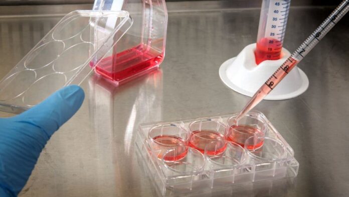
A new test could eventually help patients to avoid unnecessary surgery for ductal carcinoma in situ (DCIS), according to University of California San Diego scientists. In a research article published in Cell Reports, the La Jolla located bioengineers report that they can predict how likely a tumour is to progress to cancer based on how sticky its cells are.
The DCIS Dilemma
Ductal Carcinoma in situ (DCIS) is a “precancerous” growth of cells commonly found during routine screening for breast cancer. Doctors estimate that DCIS diagnoses make up around a fifth of new cases of breast cancer. While that sounds like a lot, in most cases DCIS doesn’t progress into full-blown cancer. As of 2010 mortality statistics for women diagnosed with DCIS were 1–2.6%. In recent years some cancer experts have even argued that classing DCIS as cancer might be doing more harm than good. This has led, they claim, to unnecessary surgeries with ensuing financial hardship and trauma for patients and their families.
On the other hand, according to some studies (albeit ones with limited sample sizes), more than a quarter of DCIS cases do eventually turn into cancer. So how to they test DCIS now?Current practice is for a pathologist to examine a DCIS biopsy under the microscope. The doctor then assigns it a grade based on whether the cells look as if they are going to become an invasive ductal carcinoma. When a growth becomes invasive, it means the tumour is spreading or metastasizing. Based on the grade allocated by the pathologist, the patient and their doctor will decide whether or not to have the DCIS surgically removed.
For many, the idea of leaving what could be a ticking time bomb in place is not something they can live with. As UCSD professor Dr. Adam J. Engler, commented to the National Institute of Health’s communications team, “There’s no good universal marker for metastasis.”
What if pathologists were to have a reliable, objective method of judging whether cells collected from a DCIS were on their way to turning cancerous?
Sorting the Good From the Bad: a DCIS test?
Dr. Engler’s team at UCSD’s Chien-Lay Department of Bioengineering is working towards finding a solution to the DCIS conundrum.
In their research paper, published in March 2025, PhD student Madison Kane and colleagues announced a method for discerning whether DCIS tumours would metastasize. The group describes in this paper how they developed a new way to calculate the proportion of cells in a biopsy that are likely to progress to invasive ductal carcinoma. The team claimed they could not only link individual cell “stickiness” to genetic cancer profiles, they have created a device that can assess a tumour’s metastatic potential. This device uses a tumour’s constituent cells’ tendency to adhere to their neighbours to predict its likeihood of metastasizing.
Engler told the NIH, “The hope with this device is that we’ll be able to say to patients, ‘You’re on the lower side of this metastatic risk spectrum’ or ‘You’re at a higher risk, so we suggest a different treatment plan,’” while this technology is in its early stages, he explains, the outlook is encouraging, “We’re not there yet. But the data is at least pointing in that direction.” We might not be seeing Engler’s lab’s invention in the clinic next week, but it does open a fascinating window into how cancerous cells behave and the novel techniques researchers are using to beat them.
Attachment Disorder?
Cancers spread through metastasis. In a nutshell, this means that a subset of cells in a tumour break free and set off to set up their own home. Cancer cells exhibit a peculiar trait: after many rounds of uncontrolled cell division and errors in DNA copying, they “forget” their identity. Once their regular jobs are forgotten, these cells start living for themselves. Over time, as they accumulate more DNA changes, the connections between the cells break down.
At a certain point, the cells don’t have a reason to stay in contact anymore. They are on their way to changing their identity and focusing on their own needs. Some cells will crawl away from the tumour to find a more hospitable, less crowded place to live, with better resources. The problem is that when they set up in their new home, they will multiply, causing a secondary tumour. Eventually the same thing will happen again when it gets too overcrowded, and the cancer will continue to travel.
Only Connect
DCIS arises from epithelial cells that line the breast’s milk ducts. Epithelial cells are generally used to cover surfaces inside and outside your body. For example, the interior of your mouth, the lining of your intestines, and the lungs. Even your skin is a type of epithelial cell.
Sheets and tubes of epithelial cells have a protective architectural role. They tend to form very strong connections that prevent cells, objects or liquid from penetrating where they shouldn’t. The attachments between the cells are so tight that only certain specialized patrolling cells, for example white blood cells, can penetrate the epithelial barrier. In epithelial cells the difference between tight connections and loose connections is big enough to measure.
The UCSD team hypothesized that they could measure how ready the cells were to drop their attachments to their neighbours. They speculated that they could then infer the probability that those cells were on the road to becoming an invasive ductal carcinoma. This meant they had three questions to ask:
How does “stickiness” correlate to a cell’s likelihood of metastasizing?
Could they reliably determine how sticky cells are in a mixed sample?
Could they use the overall “stickiness” of a sample to predict its likelihood of metastasizing?
Slow and sticky
Kane and company started by testing the relationship between a cell’s ability to form attachments and molecular markers of metastasis.
The researchers used a virus to label human breast cancer cell samples in a dish with special glowing proteins. These cells were cultivated from biopsies of a breast cancer tumour that was in the process of metastasizing. They put the cells in a dish covered in cell-friendly collagen coating and watched as the cells crawled around to find new places to grow. Cells that had a stronger ability to cling on to each other were less likely to migrate. When they did, they moved more slowly.
So was this sticky and slow situation also true in real life?
The team then injected these glowing human breast cancer cells into a mouse’s hind leg and waited six weeks. When the investigators looked for the glowing cells in the mouse’s leg, they found that the glowing human breast cancer cells had made a tumour in the mouse’s thigh. Not only was there a bright clump of glowing tumour cells in the surrounding fatty tissue, the researchers could see a few individual green glowing cells that had escaped from the main tumour.
When they collected the green glowing cells, they found that the cells that metastasized from the tumour into the fat pad were less good at forming stable attachments to other cells than the cells that stuck around in the tumour. In fact, according to the team’s adhesion studies, the cells that stuck around in the tumour were 60% stickier than the cells that escaped.
Sticky cells were less likely to metastasize.
Markers of malignancy
The researchers needed to confirm that a cell’s ability to form firm connections was an indicator of how “cancerous” a cell was. The team sorted human breast cancer cells grown in the lab into flasks based on how sticky they were. They ended up with three sets: Strongly Adherent, Weakly Adherent and unsorted control breast cancer cells. They labelled these cells with glowing proteins again and injected five mice with unsorted cancer cells, six mice with weakly adherent cancer cells and six mice with strongly adherent cells and waited six weeks.
This long incubation period gave the human breast cancer cells time to settle in and set up home. Once a primary tumour had established itself, break away groups of more advanced cancer cells set out to find new places to grow, finally taking up residence in the mouse’s lungs. This provided the researchers the chance to collect human breast cancer cells at lots of different stages along their journey to malignancy.
For each set of mice at the end of the six weeks, they looked to see whether the green cells had formed a tumour at the site of injection and examined the lungs for secondary tumours. They then collected cell samples from each of the mice to see if the tumours in the mice injected with weakly adherent (less sticky) cells were distinct from those of the mice with strongly adherent cells.
Kane then used next generation RNA transcriptome sequencing to develop gene expression profiles for the human breast cancer tumours in each set of mice.
What this means in practice is that they were able to identify almost every gene that was switched on in the cells, using high-powered DNA sequencing machines. When Kane compared the profiles of each set of cells, she soon realized that the level of a cell’s stickiness corresponded to the number of activated genes that we link to metastasis. The less sticky the cells were, the more genes to do with movement and migration they expressed. This, she and Engler inferred, meant that more sticky cells are less likely to metastasize because they are not yet running the gene expression programmes that allow them to move and migrate.
Stickiness of a cell was negatively correlated to the number of activated genes related to metastasis.
The Amazing Race
The researchers discovered that the mice injected with strongly adherent breast cancer cells developed fewer secondary tumours than mice injected with unsorted cancer cells. The mice injected with the least sticky cells developed the most secondary tumours. The stickiest breast cancer cells were the least likely to metastasize. In fact, the authors describe the most strongly adherent breast cancer cells as consistently showing “minimal metastatic activity.”
With this vital piece of evidence in hand, the team then focused on finding out whether they could use stickiness as a marker of metastatic potential. Would this relationship hold true outside the strict conditions of their sorted labelled human breast cancer cell experiments?
The bioengineers set to work devising a plan. They acquired seven more human breast cancer cell cultures created from different tumours and used adhesion assays to determine how sticky each one was. They created a race track/assault course, using collagen hydrogel coatings that mimic the texture of a breast cancer tumour. The researchers measured how fast cells in each of the tumour lines could crawl across the collagen matrix. Then they used parallel-plate flow chambers to test how sticky the cells were.
The idea was to rate cell stickiness, based on how much force they needed to use to dislodge the cancer cells from a fibronectin coating on a dish. They put the cells in the dish; they settle and cling on to the fibronectin matrix, mimicking how they would grab hold of neighbouring cells in their usual environment. Then liquid was washed across the surface of the dish and the investigator counted how many cells were knocked off. Depending on how many cells were dislodged, they gave the cell sample an overall stickiness rating.
Sticking Points Show the Way
Kane and Engler developed a parallel-plate flow chamber that could measure distinct levels of stickiness by varying the amount of force applied over different parts of the chamber. This allowed Kane to assess not just whether the cells were sticky or not sticky, but increments of stickiness. This incremental measure is the key to the technique.
The team trialled the seven cell samples against each other to see which cancer line had the fastest-moving cells, and whether each cell sample’s stickiness was consistently inversely proportional to their speed. In every sample the team found that the more active, wandering cells were less sticky. Another point in favour of the idea that sticky cells equals a less invasive tumour.
They could reliably measure how sticky a mixture of cells was at multiple points along a scale from weakly adherent to strongly adherent using their adapted parallel plate flow chamber.
Translating the tech
The question now: if they could link the stickiness of individual cells to metastatic potential, could they do it with a collection of cells? It’s all very well to spot a single super-speedy cell in a dish, but to get this technology tucked into every pathology lab, it would need to be able to rate an assortment of jumbled up cells taken from a breast cancer biopsy. In that sample, you would find a range of different cells at different stages of malignancy and you wouldn’t have the helpful glowing protein labels guiding you to separate cancer cells from non-cancer cells. Could they use their improved parallel-plate flow chamber to distinguish specimens with high metastatic potential based on the proportion of weakly adherent cells they contain?
Kane and colleagues used the data they collected in the previous adhesion assays to set a cut-off limit for the amount of force needed to dislodge a predetermined ratio of cells if they had the characteristics of a cell ready to metastasize. They gathered samples from mice injected with labelled human cancer cells but this time they dropped the cells into the parallel plate flow chamber. They discovered that non-cancerous cells were able to form strong connections to the fibronectin coatings, whereas cancerous cells became less adhesive as their inclination to metastasize increased. The team set up a series of experiments to test whether they could predict a sample’s invasive potential by checking the average stickiness of the cells.
They used their existing data to set stickiness thresholds for distinct degrees of metastatic potential. That is, they were able to make a scale of stickiness and mark increments along it that signified how likely it was that the tumour would metastasize. The group tested their rating scale by mixing samples of cells taken from various breast cancer cell lines. Somebody would make a mystery sample by mixing cells collected from different cell lines with different metastatic prospects. Another member of the team would run the cell mix through the modified parallel plate lateral flow chamber to generate an adhesion profile of the sample. They would then use their stickiness rating tool to see if they could identify which cell lines had been chosen and in what proportions.
From in vitro to in vivo
So Kane and company had tested their stickiness scores on cells grown in the lab and carefully chosen cell cocktails, but would this work on messy mixtures with non-cancerous cells and real life complications?
The team returned to their mouse model. They injected mice with strongly adhesive human breast cancer cells with high metastatic potential and mice injected with weakly adhesive cells with low potential for invasion. Then, after the usual waiting time of a few weeks, they collected samples from the original injection site—the primary tumour, and from secondary tumours in the lungs. The team then used their modified parallel plate flow chamber assay to test the cells. Could they distinguish between samples from the mice injected with cells of low metastatic potential or high metastatic potential? They could indeed. They were able to correctly guess that a tumour sample had come from one set of mice or the other based on how sticky the cells in each sample were.
Finally they tested samples taken directly from biopsies from women diagnosed with ductal carcinoma in situ to see whether their predictions based on stickiness were consistent with observations made by the pathologist.
In each case, the team was able to distinguish between specimens with a high risk of metastasis or a lower risk, based on the stickiness of the cells determined by their improved parallel plate fluid chamber experiment.
DCIS under the spotlight
The researchers showed that they could match a cell’s adhesive capabilities to its motility and to its likelihood of expressing genes involved in metastasis. They demonstrated a novel approach to measure and objectively determine a mixed cell sample’s adhesion profile. Finally, the investigators found that they could use this measure of adhesion to predict how likely a tumour was to metastasize.
While this project is relatively early in its journey, Engler’s team has come up with a truly novel way to study how tumours develop towards malignancy. This will be especially useful to DCIS researchers still getting to grips with the characteristics of the condition.
Is DCIS cancer? Is it even pre-cancer? How soon in its progress does it become clear whether the tumour will eventually become invasive?
Thorough testing, evaluation and more research and development will be needed before the FDA can approve this method is ready for widespread use, but these encouraging results will light the road on the way to a more informed standard of care for DCIS.
Bibliography
Deep learning for predicting invasive recurrence of ductal carcinoma in situ: leveraging histopathology images and clinical features – eBioMedicine. Accessed June 5, 2025. https://www.thelancet.com/journals/ebiom/article/PIIS2352-3964(25)00194-X/fulltext
Kane MA, Birmingham KG, Yeoman B, et al. Adhesion strength of tumor cells predicts metastatic disease in vivo. Cell Reports. 2025;44(3):115359. doi:10.1016/j.celrep.2025.115359
Kerlikowske K. Epidemiology of Ductal Carcinoma In Situ. J Natl Cancer Inst Monogr. 2010;2010(41):139-141. doi:10.1093/jncimonographs/lgq027
Morrow M, Barrio AV. Is It Time to Abandon Surgery for Low-Risk DCIS? JAMA. 2025;333(11):946-947. doi:10.1001/jama.2024.26723
Partridge AH, Hyslop T, Rosenberg SM, et al. Patient-Reported Outcomes for Low-Risk Ductal Carcinoma In Situ: A Secondary Analysis of the COMET Randomized Clinical Trial. JAMA Oncology. 2025;11(3):300-309. doi:10.1001/jamaoncol.2024.6556
Rubin R. Experts Are Debating Whether Some Cancers Shouldn’t Be Called That. JAMA. 2025;333(7):556-558. doi:10.1001/jama.2024.27250


